Circuit bases for sensory information processing in the brain
The mammalian nervous system is composed of enormous numbers of neurons, but how do these cells take on diverse fates and organize and array themselves during development? How is a functional circuit formed during? We study neural circuits for olfaction (see our recent review), somatosensation, audition, and interoception (gut chemical senses) as model systems.
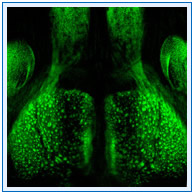
We use in vivo two-photon calcium imaging to study the dynamics of sensory information processing (see a press release and a blog post on our recent paper). We also employ mathematical simulation to understand the circuit dynamics. Circuit bases are now being studied by the light microscopy-based connectomic approaches, incorporating tissue clearing techniques developed recently in our laboratory (e.g., SeeDB, SeeDB2). Our goal is to understand the developmental mechanisms of the functional neuronal circuitry.
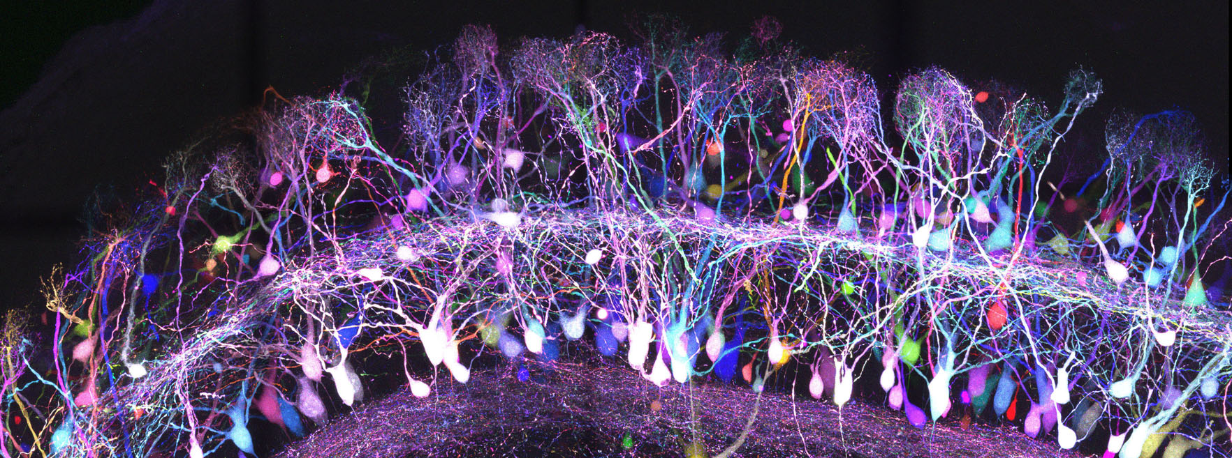
Apart from biology, we are also trying to develop new tools in neuroscinece, particularly in connectomics. We believe that new tools reveal new questions and new fields in biology. We have previously developed tissue clearing methods enabling large-scale 3D analyses of neuronal circuits. We are trying to develop new strategies in light microscopy-based connectomics.